The 20 Best Microscope Photos from the 2017 Nikon Small World Contest
The Nikon Small World Photomicrography Competition celebrates the beautiful, weird, wonderful, and microscopic things in our world through photos captured using a light microscope. Now in its 43rd year, the competition continues to impress.
“This year’s winners not only reflect remarkable research and trends in science, but they also allow the public to get a glimpse of a hidden world,” says Eric Flem, Communications Manager, Nikon Instruments, “This year’s winning photo is an example of important work being done in the world of science, and that work can be shared thanks to rapidly advancing imaging technology.”
So, without further ado, here are the 20 amazing images that placed in the competition this year:
1st Place

Photo by Dr. Bram van den Broek, Andriy Volkov, Dr. Kees Jalink, Dr. Reinhard Windoffer & Dr. Nicole Schwarz.
Immortalized human skin cells (HaCaT keratinocytes) expressing fluorescently tagged keratin.
2nd Place
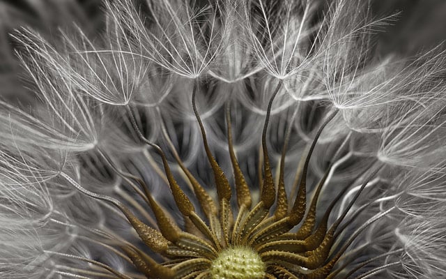
Photo by Dr. Havi Sarfaty.
Senecio vulgaris (a flowering plant) seed head.
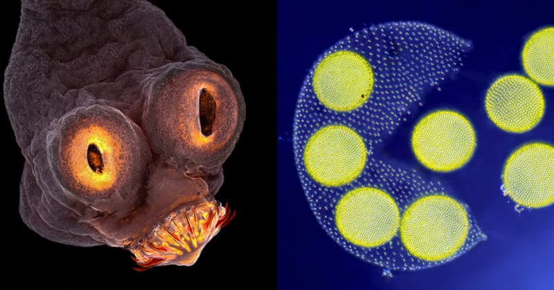
3rd Place
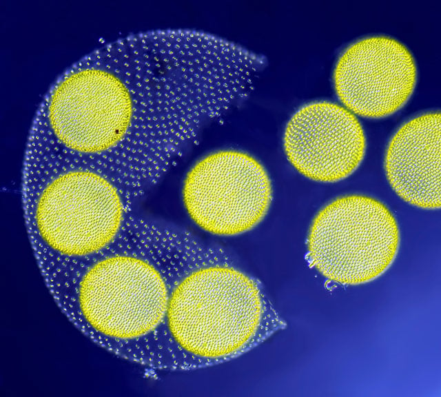
Photo by Jean-Marc Babalian.
Living Volvox algae releasing its daughter colonies.
4th Place
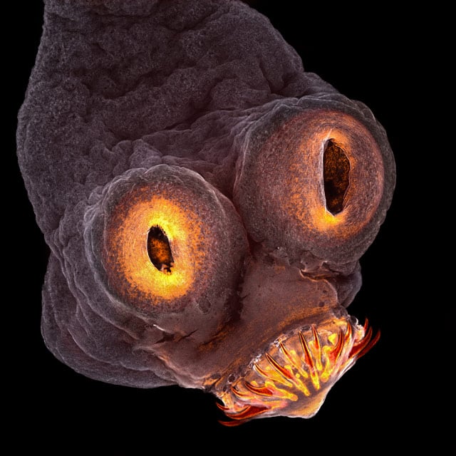
Photo by Teresa Zgoda.
Taenia solium (tapeworm) everted scolex.
5th Place
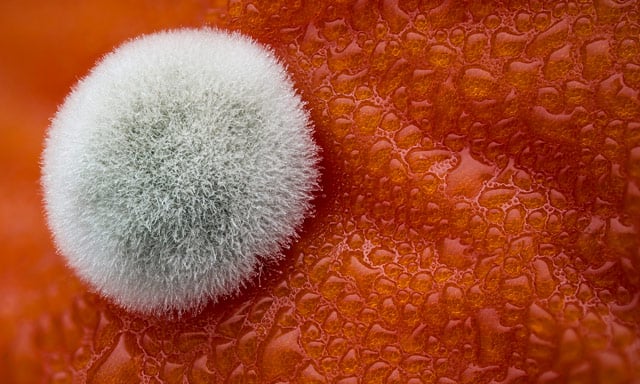
Photo by Dean Lerman.
Mold on a tomato.
6th Place
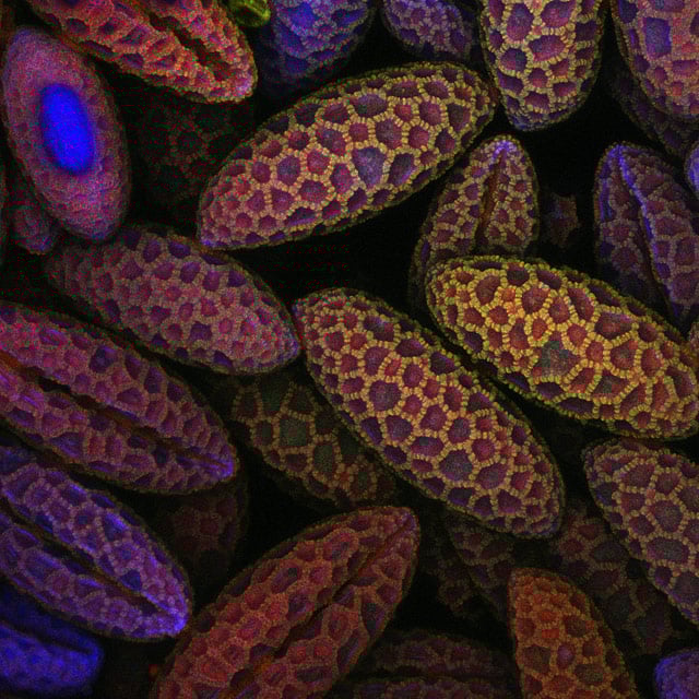
Photo by Dr. David A. Johnston.
Lily pollen.
7th Place
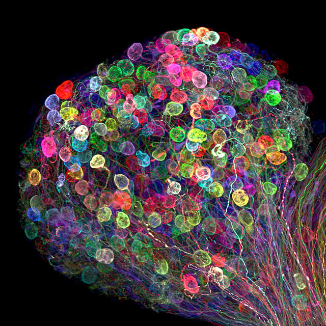
Photo by Dr. Ryo Egawa.
Individually labeled axons in an embryonic chick ciliary ganglion.
8th Place
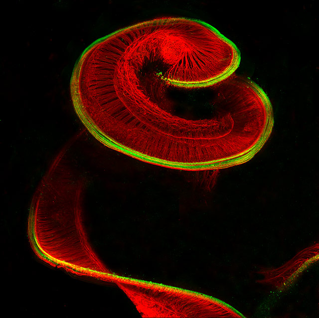
Photo by Dr. Michael Perny.
Newborn rat cochlea with sensory hair cells (green) and spiral ganglion neurons (red).
9th Place
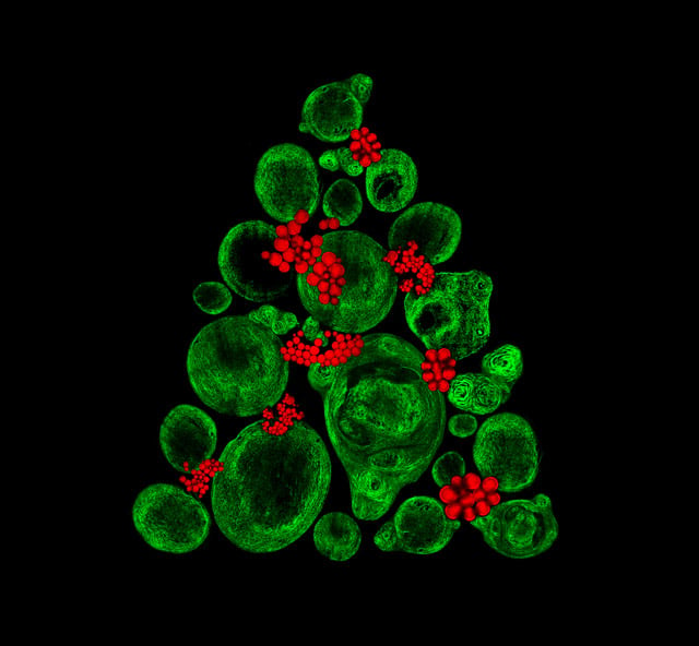
Photo by Catarina Moura, Dr. Sumeet Mahajan, Dr. Richard Oreffo & Dr. Rahul Tare.
Growing cartilage-like tissue in the lab using bone stem cells (collagen fibers in green and fat deposits in red).
10th Place
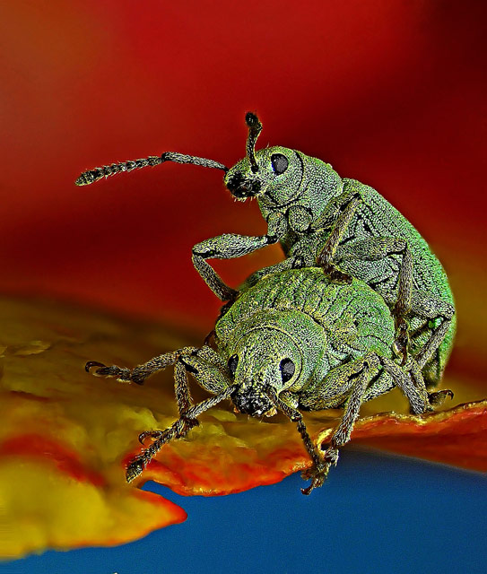
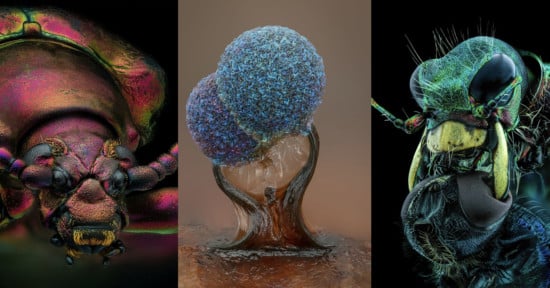
Photo by Dr. Csaba Pintér.
Phyllobius roboretanus (weevil).
11th Place
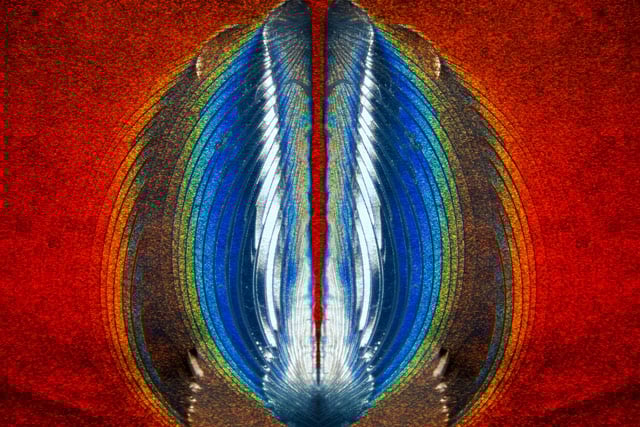
Photo by Steven Simon.
Plastic fracturing on credit card hologram.
12th Place
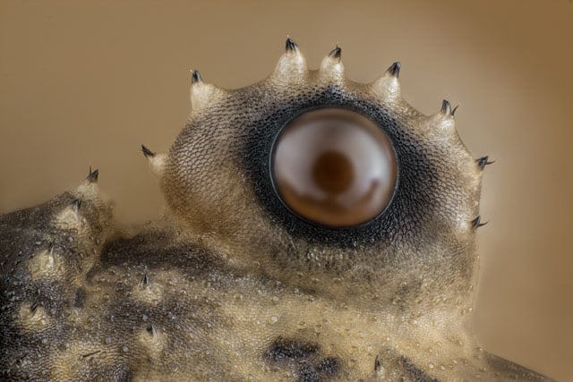
Photo by Charles B. Krebs.
Opiliones (daddy longlegs) eye.
13th Place
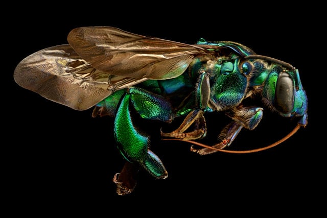
Photo by Levon Biss.
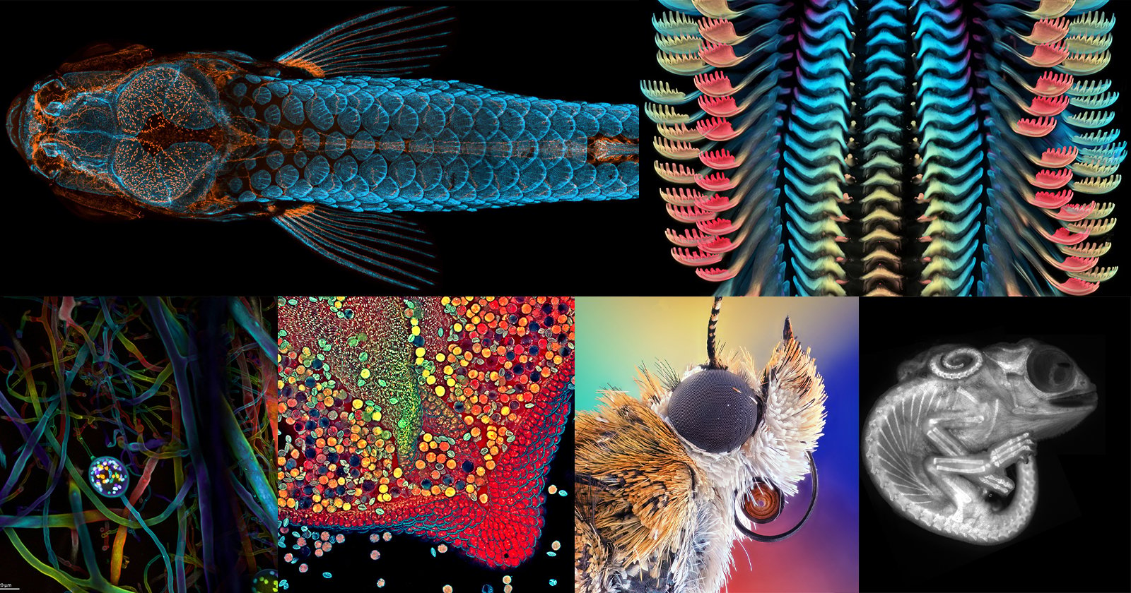
Exaerete frontalis (orchid cuckoo bee) from the collections of the Oxford University Museum of Natural History.
14th Place
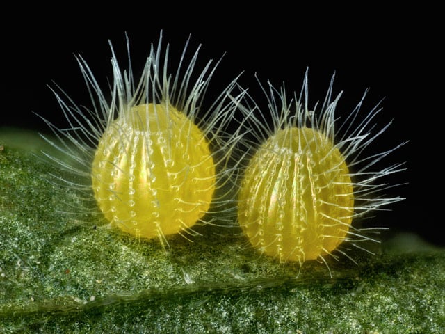
Photo by David Millard.
Common Mestra butterfly (Mestra amymone) eggs, laid on a leaf of Tragia sp. (Noseburn plant).
15th Place
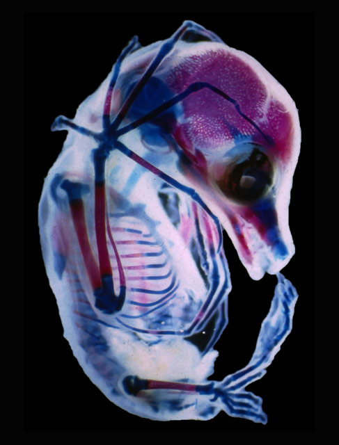
Photo by Dr. Rick Adams.
3rd trimester fetus of Megachiroptera (fruit bat).
16th Place
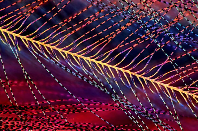
Photo by Marek Miś.
Parus major (titmouse) down feather.
17th Place
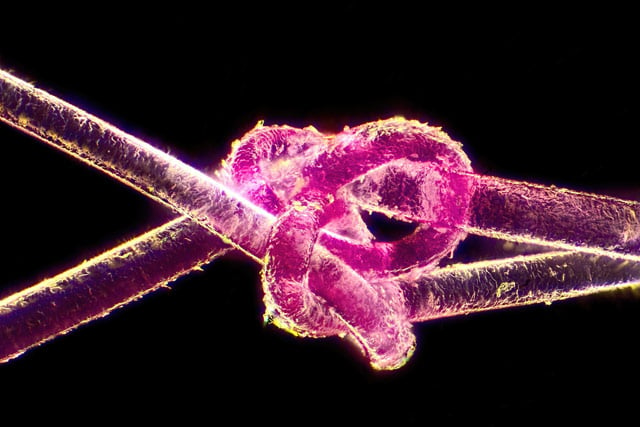
Photo by Harald K. Andersen.
Dyed human hair.
18th Place
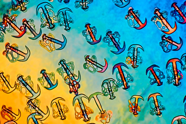
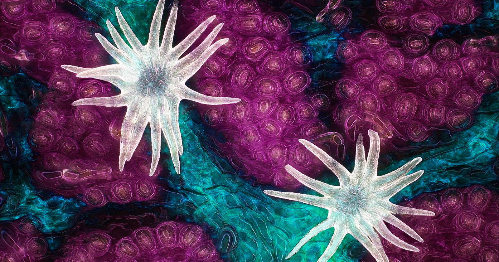
Photo by Christian Gautier.
Synapta (sea-cucumber) skin.
19th Place
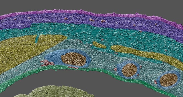
Photo by Dr. Dylan Burnette.
Embryonic body wall from a developing Mus musculus (mouse).
20th Place
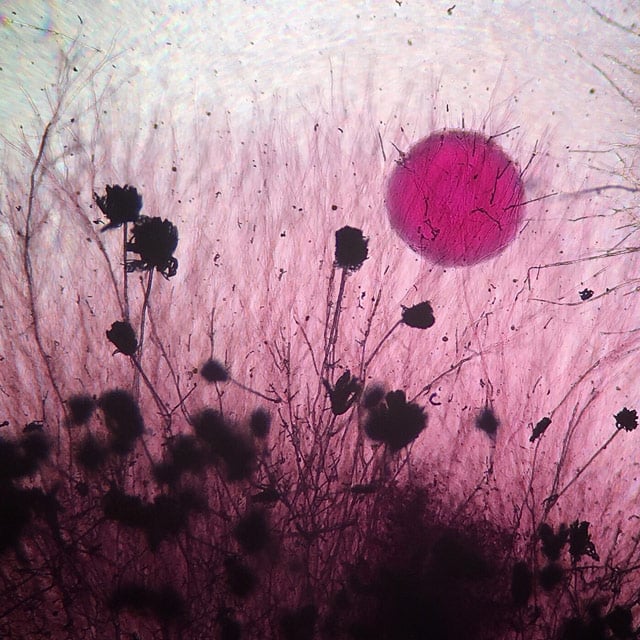
Photo by Tracy Scott.
Aspergillus flavus (fungus) and yeast colony from soil.
You can see other awarded entries on the Nikon Small World website.