by
Kathy Mahdoubi, Senior Correspondent | July 08, 2009
This report originally appeared in the June 2009 Nuclear Medicine issue of DOTmed Business News
Some might say the status quo of orthopedic imaging remained relatively undisturbed for the better part of the 1980s, and perhaps even the '90s, but in a matter of just a few years, a relatively new hybrid of molecular imaging and computed tomography, SPECT/CT, has emerged and quickly become the new gold standard for bone imaging and the pathology of skeletal disease.
The combination of single photon emission computed tomography (SPECT), and X-ray computed tomography (CT) has been championed by cardiologists since its inception, but the modality has also gained a great deal of ground in orthopedic imaging and bone pathology. SPECT is a highly sensitive technology that captures functional changes associated with a wide variety of bone and joint tissue abnormalities, including inflammation, infection, benign and malignant lesions, and bone metastases. SPECT is also considered a significantly more affordable alternative to PET imaging.
Standalone modalities have limitations
"The problem with general X-ray is that it's not volumetric, and X-ray and CT can't pick up inflammation or infection," says Dominic Smith, Vice President of Nuclear Medicine Global Marketing for Philips Healthcare. "That's why the combination of SPECT and CT is very good for orthopedics."
Magnetic resonance technology is also problematic when applied to orthopedic procedures, particularly if patients have undergone hip or knee replacement or arthroscopic surgery and have metal implants or prosthetics in place, says Smith.
Upon first examination, a practicing physician treating a patient complaining of unexplained bone pain may decide to order an X-ray, "but when a patient is having bone pain and nothing appears in radiographic imaging, the next step is a bone scan," says Dr. Richard Myers, Chairman of the nuclear medicine department at Radiological Associates of Sacramento, a CA-based radiology practice specializing in diagnostic imaging, nuclear medicine and radiation oncology.
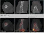
Osteosarcoma of the knee.
0.33 mm thick isotropic
CT slices acquired from
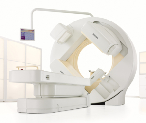
a Philips Brightview XCT
SPECT systems are scintigraphic, meaning they employ gamma camera technology to detect gamma rays emitted by injected radioisotopes localized at areas of increased bone metabolism. Unlike planar gamma camera imaging, SPECT uses multi-planar reconstruction algorithms to yield datasets that can formulate 3-D images of bone and joint tissue.