A Two-Headed Shark and Other X-Rayed Beauties at the Smithsonian
Sandra Raredon’s x-rays of fish specimens are critical records for scientists studying various species. And, as works of art, they are breathtaking
/https://tf-cmsv2-smithsonianmag-media.s3.amazonaws.com/accounts/headshot/megan.png)
/https://tf-cmsv2-smithsonianmag-media.s3.amazonaws.com/filer/a6/dc/a6dc79e7-5a30-452c-8e2f-e6983e60b0ed/usnm_39499_mustelus_radiograph_two_headed_body.jpg)
The National Museum of Natural History maintains the largest fish collection in the world. Most of the 4 million specimens, including adult fish, eggs, larvae and juveniles, are stored in jars of ethanol, which fill six massive rooms at the Smithsonian’s Museum Support Center in Suitland, Maryland.
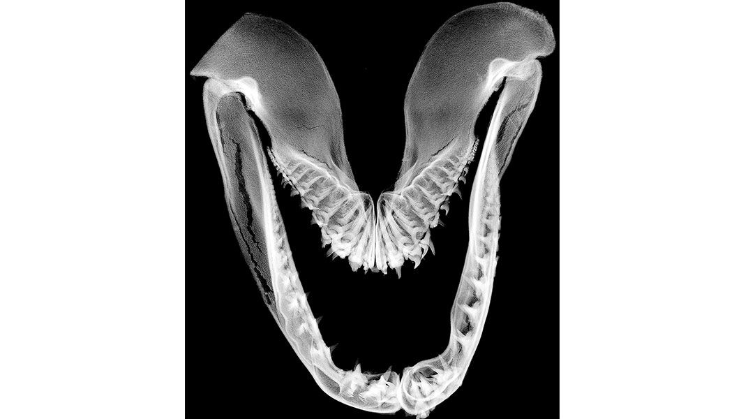
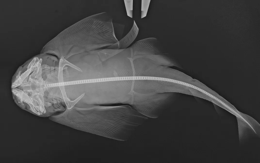
If you are lucky enough to get an invitation to the facility's wet pod, a staffer might treat you to what is called the "Oh my" collection—a sampling of the fish division's greatest hits, including a piranha, a lionfish, some fish collected by Teddy Roosevelt, gorgeous chimeras and an electric eel. But perhaps the most shocking specimen is the two-headed shark.
"You don't see those everyday," says Sandra Raredon, a museum specialist. When she x-rayed the smooth-hound seen above, a larval specimen about six inches long, she found two separate vertebral columns.
A 27-year employee, Raredon helps to maintain the expansive "fish library." The shelves contain about 75 percent of the more than 32,000 known fish species. The oldest specimens were collected in the mid-1800s, and scientists add thousands more each year, knowing that they will be preserved and shared with the scientific community.
One of Raredon's duties is to x-ray each of the specimens. She removes a one- to two-foot shark from its container, places it on her x-ray machine's digital tablet and applies an exposure of about 40 kilovolts for five to ten seconds. The tablet captures x-rays that pass through the fish and creates a digital image of its internal structure on Raredon's computer monitor.
For ichthyologists, or scientists who study fish, having access to the skeletons of fish is particularly important. When scientists find what they think might be a new species, for instance, they count the specimen’s vertebrae and fin spines and examine its teeth and the structure of its caudal fin, or tail. Then they compare those numbers and observations with known species found in fish collections. Comparing skeletons can also help scientists figure out how groups of fishes are related and how fish have evolved over time.
One way to get at a skeleton, especially a large one, is through dissection. There are more than 4,000 dry fish skeletons in the museum’s collection. Another method, used on small fish that would curl up if dried, is called “clearing and staining.” The fish is soaked in trypsin, a digestive enzyme, to clear the flesh away, and the cartilage is stained blue while the bone is stained red. Stored in glycerin afterward, these are often referred to as “wet” skeletons; the museum has more than 5,000. But, x-rays, which have been used to study fish since shortly after the form of radiation was discovered in 1895, are especially noninvasive, in that they don't alter the specimen.
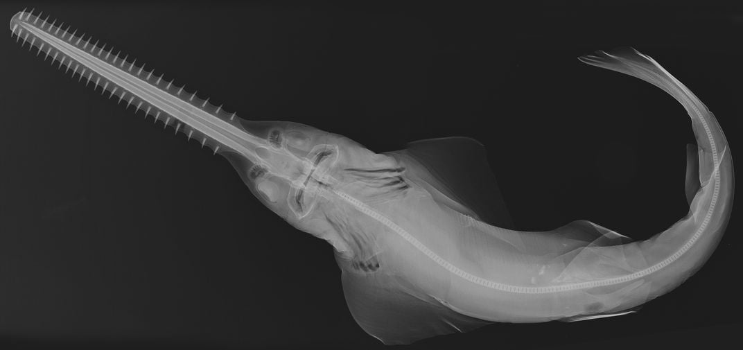
Raredon is gradually x-raying her way through the collection, giving first priority to “types,” or the original specimens from which species were identified and named; old specimens that are degrading; and fish that resident and visiting scientists request she x-ray for their research. In total, she has taken more than 11,000 x-rays of specimens, including sawfish, hammerheads and angel sharks. She logged the first 10,000 using a chemical film-developing process. In 2001, she switched to taking digital x-rays when the museum purchased its first digital radiographic machine. Whereas a conventional x-ray requires about 30 minutes to develop and longer to dry, the digital versions are instantly ready to study and send to researchers around the world. Not to mention, scientists can zoom in or invert the black-and-white x-rays to see a fish’s bone structure more clearly. "These x-ray machines are just as important as a microscope in our work," Raredon explains.
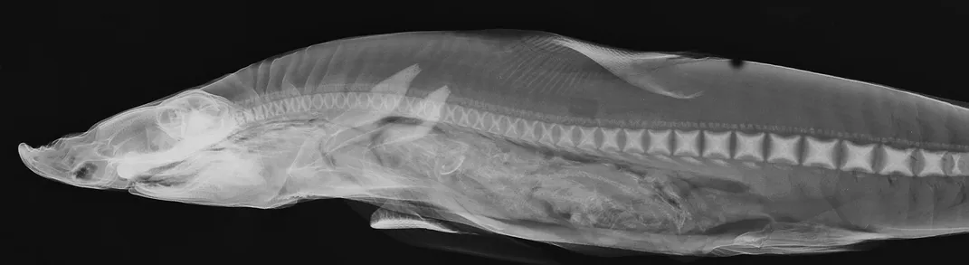
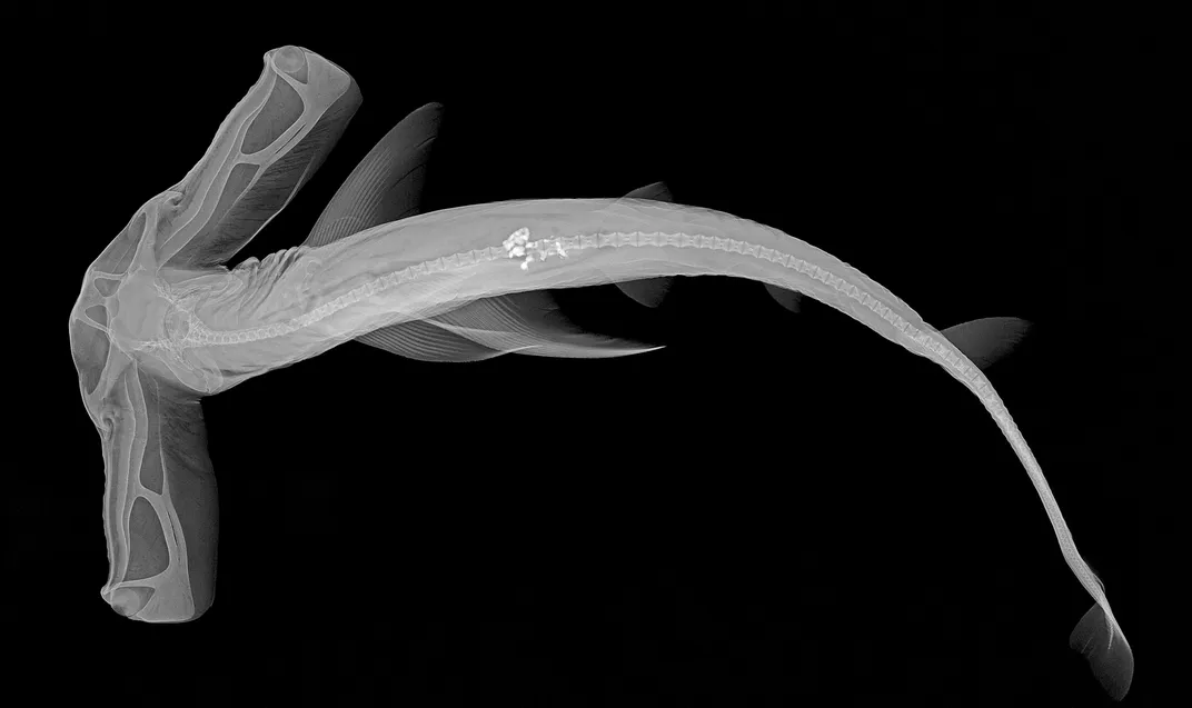
Occasionally, when taking x-rays of specimens, Raredon notices last suppers in the fishes' guts. She found an animal in the belly of a whitecheek shark, for instance. "When you blow it up a little bit, you can see another fish inside," she says of a lateral view of the shark (shown in the gallery, above). "You can see a long series of vertebral bones in there." In an x-ray of a winghead shark (also shown), there are bright white remains in its mid-section. "Could be a clam or something," says Raredon.
There is certainly an artistry to the x-rays. While they serve scientific purposes, they can also be appreciated for their delicate aesthetics. Raredon helped to compile a selection of the eye-catching x-rays into Ichthyo: The Architecture of Fish, a book published in 2008, and "X-Ray Vision: Fish Inside Out," a Smithsonian exhibition traveling to museums, aquariums, libraries and universities around the country.
Raredon is partial to her x-rays of stingrays, but also has a soft spot for the sharks.
"Look at the hammerheads," she says. "They are very dangerous animals, but then when you look at them like this, white on black, everything is symmetrical, and they are very graceful. They are beautiful."
/https://tf-cmsv2-smithsonianmag-media.s3.amazonaws.com/accounts/headshot/megan.png)